
When someone is diagnosed with cancer, a common question is: What makes my cancer unique?
At MUSC Hollings Cancer Center, researchers are working to answer that question in a new way – by studying tiny sugar molecules that coat our cells.
These molecules, called glycans, may hold powerful clues about how a cancer behaves, how it interacts with the immune system and how it might respond to treatment.
Through a unique partnership between MUSC and Bruker, a global scientific instrumentation company whose technologies are widely used in biomedical research and diagnostics, the Bruker–MUSC Center of Excellence in Clinical Glycomics is turning this advanced science into tools that could one day guide more personalized cancer care.
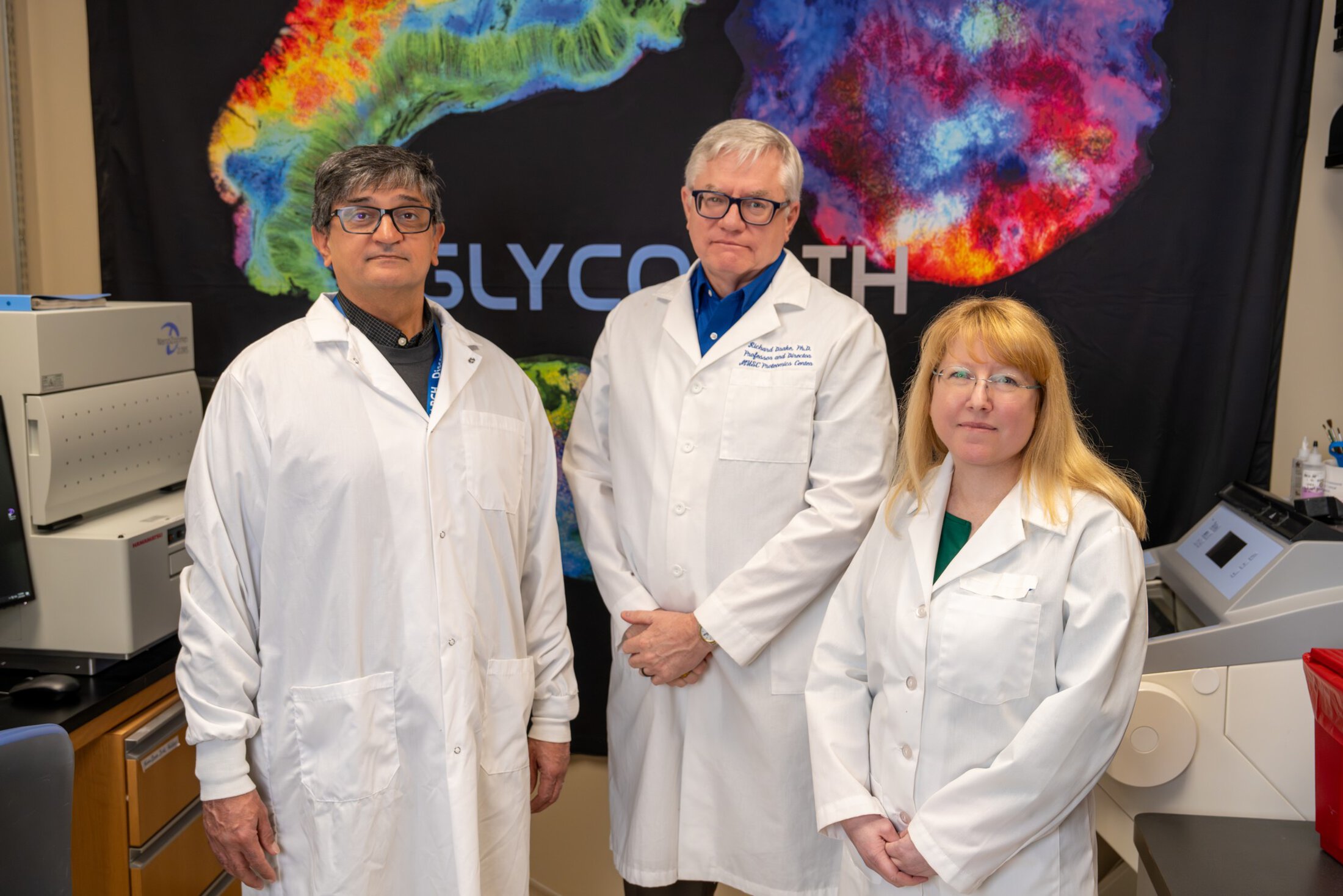
The center grew from decades of collaborative work by Richard Drake, Ph.D.; Peggi Angel, Ph.D.; and Anand Mehta, Ph.D., in the Hollings Developmental Cancer Therapeutics (DCT) Research Program. Their shared expertise in glycosylation and cancer biomarkers laid the foundation for the program.
At the core of their work is an understanding of how sugars attached to proteins can reveal critical information about disease.
“The blood and tissue samples each patient provides all have sugars attached to them that reflect their condition – we have developed new ways to rapidly visualize and identify these sugars,” said Drake.
Why glycans matter
Cancer often leaves subtle clues long before it spreads or stops responding to treatment – but many of those clues remain hidden. At the center, researchers are working to make those signals visible.
Glycans are complex sugar structures that cover the surface of cells and proteins throughout the body. They are not the same as sugar in our diets. Instead, they act like molecular “tags” that help cells to communicate and function properly.
If something is going to matter clinically and help patients, it has to fit into real-world timelines. You can’t wait weeks for an answer.
Unlike genes, which follow a fixed template, glycan patterns shift depending on a cell’s metabolism, health and environment. When cells become cancerous, those patterns can change dramatically.
“Cancer is a metabolic disease that alters the way cells use energy,” Drake explained. “When you change how a cell uses fuel like glucose or glutamine, you change the glycans. Those changes show up on tumor cells, immune cells and proteins circulating in the blood.”
As such, glycans may serve as fingerprints of disease – subtle biological markers that reveal how aggressive a tumor is or how it is interacting with the immune system.
From pathology slides to spatial maps
Historically, glycans have been hard to study. Scientists knew they were important but lacked practical ways to study them in patient samples. The MUSC team set out to change that.
Using Bruker’s advanced mass spectrometry platforms, the center has developed new enzymatic and imaging-based assays that can analyze glycans directly from patient samples, including standard pathology tissue collected during surgery or biopsy, and generate results in hours instead of weeks.
“That was a turning point,” Drake said. “If something is going to matter clinically and help patients, it has to fit into real-world timelines. You can’t wait weeks for an answer.”
One of the center’s signature advances is imaging mass spectrometry. This technology creates detailed spatial maps showing where specific glycans appear within a tumor and in surrounding tissue. These glycan maps can be layered with traditional pathology findings and genetic testing results to build a more complete picture of a patient’s cancer.
“All these methods start with tissue that came out of pathology,” Drake said. “That makes it possible to combine the information and see the tumor in a more integrated way.”
The center’s work extends beyond tumor cells as well. For instance, Angel led a recent study showing that certain glycans are linked to triple-negative breast cancer survival and may be detected at much earlier stages within the tissue microenvironment. She has subsequently developed new approaches to studying changes in the extracellular matrix – the structural framework surrounding cells that tumors invade as they grow and spread. Changes in this environment influence how cancer progresses and how it responds to therapy.
By analyzing glycan patterns and the tumor environment, the researchers hope to understand more fully how cancers evolve and potentially uncover new treatment targets.
A new window into improved immunotherapy
The center has also expanded its focus to immune cells – a critical frontier as immunotherapies and CAR-T cell treatments become more common – through close collaboration with the Center for Cellular Therapy, led by Shikhar Mehrotra, Ph.D.
Immunotherapies harness the body’s immune system to fight cancer, but they do not work for everyone. Some patients respond dramatically, while others see little benefit or experience serious side effects. Glycans may help to explain why.
“The immune system is essentially a glycan-sensing machine,” Drake said. “Immune cells are covered in lectins, which are proteins that bind sugars. They’re constantly reading glycans on the surface of other cells.”
Every person’s cancer is different. These tools give us additional ways to understand those differences at a fundamental level and to use that knowledge to guide more personalized care.
Mehta’s group recently developed a new method to study patients’ immune cells. By adapting imaging techniques to study individual cells, researchers can analyze glycans, lipids and other metabolites at extremely high resolution. This approach offers new insight into why some patients respond to immunotherapy and others do not. That knowledge could one day help doctors to match patients with treatments more precisely.
Researchers at the center are also exploring how glycan patterns on tumors and immune cells influence immune recognition and treatment response. The findings could lead to better companion diagnostics and improved patient selection for immunotherapy trials.
Impact close to home – and beyond
For patients in South Carolina, the center’s presence offers access to technologies that currently exist nowhere else.
“Having this capability here truly matters,” Drake emphasized. “It means patients treated at Hollings can benefit from cutting-edge tools – not years down the road – but now.”
The center also has a growing international footprint. Each year, researchers from around the world travel to MUSC for hands-on training, bringing their own samples and leaving with data and expertise they can apply at their home institutions.
While much of the center’s work is still in the research and clinical trial phases, the long-term goal is clear: to integrate glycan analysis into the standard practice of precision medicine. This could eventually lead to better tools to predict how aggressive a tumor might be, more accurate ways to match patients with immunotherapies or new blood-based tests to monitor disease.
As the center enters its second five-year phase, priorities include integrating glycan-based assays into clinical trials, expanding blood-based biomarkers and strengthening collaborations in cell therapy and immuno-oncology across MUSC.
“Every person’s cancer is different,” Drake said. “These tools give us additional ways to understand those differences at a fundamental level and to use that knowledge to guide more personalized care.”
Researchers in this story
Peggi Angel, Ph.D.
Dr. Peggi Angel's research is focused on understanding the spatial systems biology of human health – how molecular interactions change due to external, endogenous environmental and mechanical forces in normal development and in disease. The primary analytical research in her lab is focused on developing new approaches for deeper single cell sequencing of collagen structures, targeting signaling components of fibrotic deposition, and the application of these methods for human disease prognosis and diagnosis. The main biological research focus of her group is understanding how spatial changes in translational and post-translational collagen regulation contributes to breast cancer initiation and metastasis and impacts on cancer risk. She is the inventor of the spatial method targeting collagen on formalin-fixed, paraffin-embedded tissues. The approach generates data that may be used not only to understand stroma regulation within the tumor microenvironment and circulating serum. Collagen biosignatures also be used as a predictive tool developing signatures that differentiate pathologies, patient status, and therapeutic response. She has published several papers using this method in various cancerous tissues. Her lab is focused on leveraging this approach to understand disparities in breast cancer, colorectal cancer, and outcomes in hepatocellular carcinoma.

Richard Drake, Ph.D.
Core Leader, DDRCC Proteomics Core
Professor, Department of Pharmacology & Immunology
SmartState Endowed Chair in Proteomics
Since arriving at MUSC in 2011, I have strived to expand translational research opportunities within my department and throughout MUSC by combining clinical research, biorepository, and molecular pathology resources with an extensive biomedical proteomics and small molecule mass spectrometry facility. My primary research focus is in identifying glycoproteins, glycans, and glycolipids using human and animal model tissues and biofluids.
My laboratory developed the founding MALDI imaging mass spectrometry methodology to allow researchers worldwide to access N-glycosylation from the tissue microenvironment in both formalin-fixed, paraffin-embedded and frozen tissues. The glycan tissue maps serve as guides to target tumor-localized glycoproteins for proteomic analysis, as well as provide molecular determinants for histopathology applications. This technology has recently been adapted and funded to develop tools to analyze cell lines, immune cells, and biofluids directly on slides. New research efforts are focused on developing new methods for investigating O-glycan, glycogen, and glycosaminoglycan oligomers, as well as expanding prior glycosphingolipid workflows.
Anand S. Mehta, Ph.D.
Endowed Chair, SmartState Chair - Proteomic Biomarkers
Professor, College of Medicine
A large part of my laboratory is focused on understanding and developing diagnostic methods and treatments for hepatocellular carcinoma (HCC), a primary cancer of the liver that kills close to 1 million people every year. Our lab was one of the first to perform total serum glycan analysis for biomarker detection and one of the first to perform serum glycoproteomics. Through this we identified over 30 serum glycoproteins with increased levels of fucose in those with HCC.
Hayley Kamin
Communications Manager
Hayley Kamin is the communications manager for the Hollings Cancer Center Communications and Marketing team, having joined the team in 2025 after three years as a communications specialist at the National Institutes of Health (NIH). As a science communicator with a Ph.D. from the University of Florida, she has extensive experience translating complex research into clear, engaging content. Her career has included roles at the NIH’s National Institute of Mental Health and the American Psychological Association, where she led content development and editorial strategy, developed science and health communications and worked with researchers and clinicians to strengthen public understanding of research.