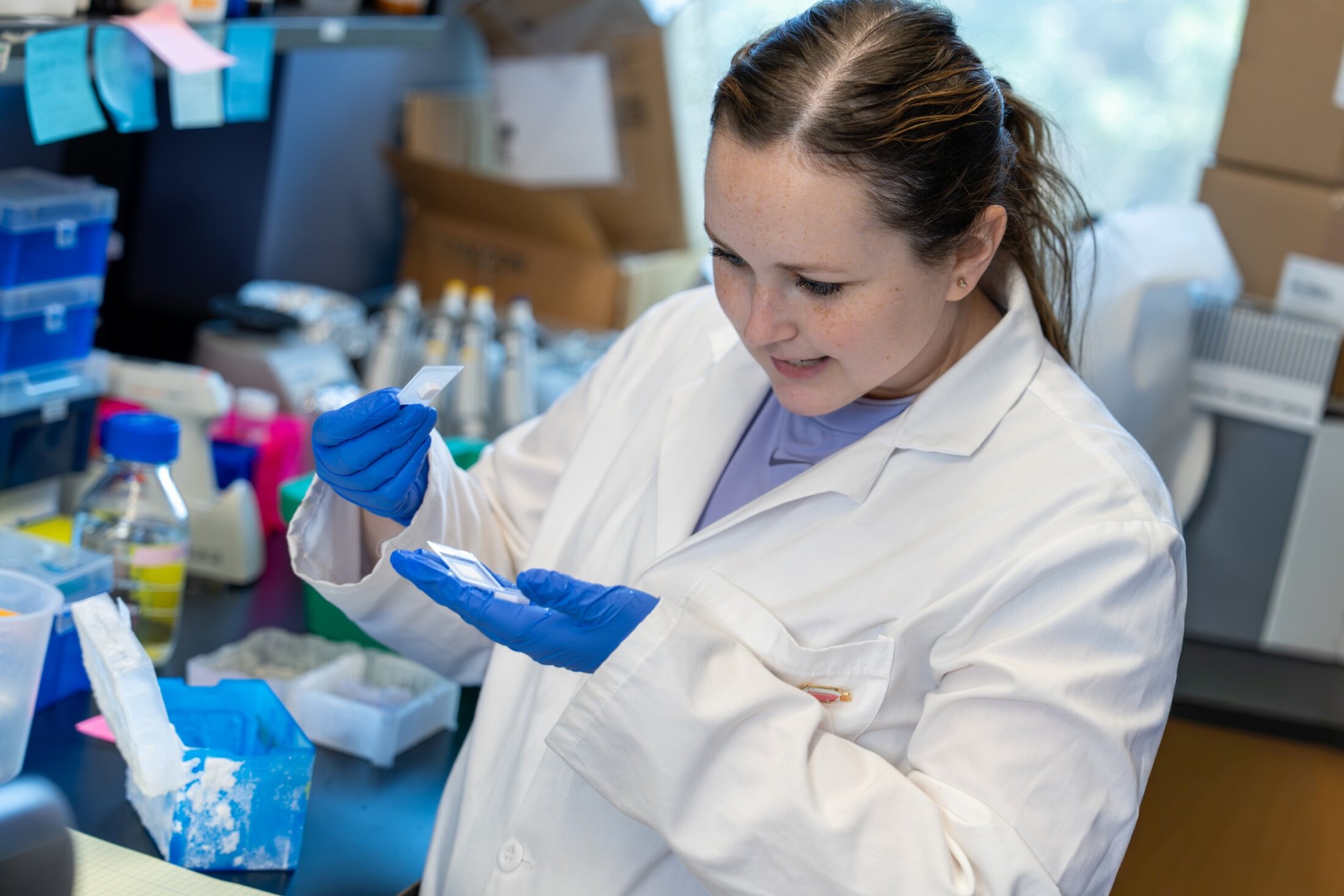
Postdoctoral fellow Allison Reno, Ph.D., uses inspiration from the world around her and from her materials science background to build better models of sarcoma. Video by Clif Rhodes
The Great British Bake Off offers drama, baking tips and – for one viewer in South Carolina – inspiration for creating 3D models that can be used to test cancer drugs.
“I borrow techniques sometimes,” said Allison Reno, Ph.D. “I see them do things that work very well for chocolate or gelatin in baking, like temperature changes. Similar principles apply to hydrogel polymerization. We can induce polymerization through rapid temperature changes, acid-based reactions and/or gentle mixing. We use a combination of those three to create a really, really strong gel.”
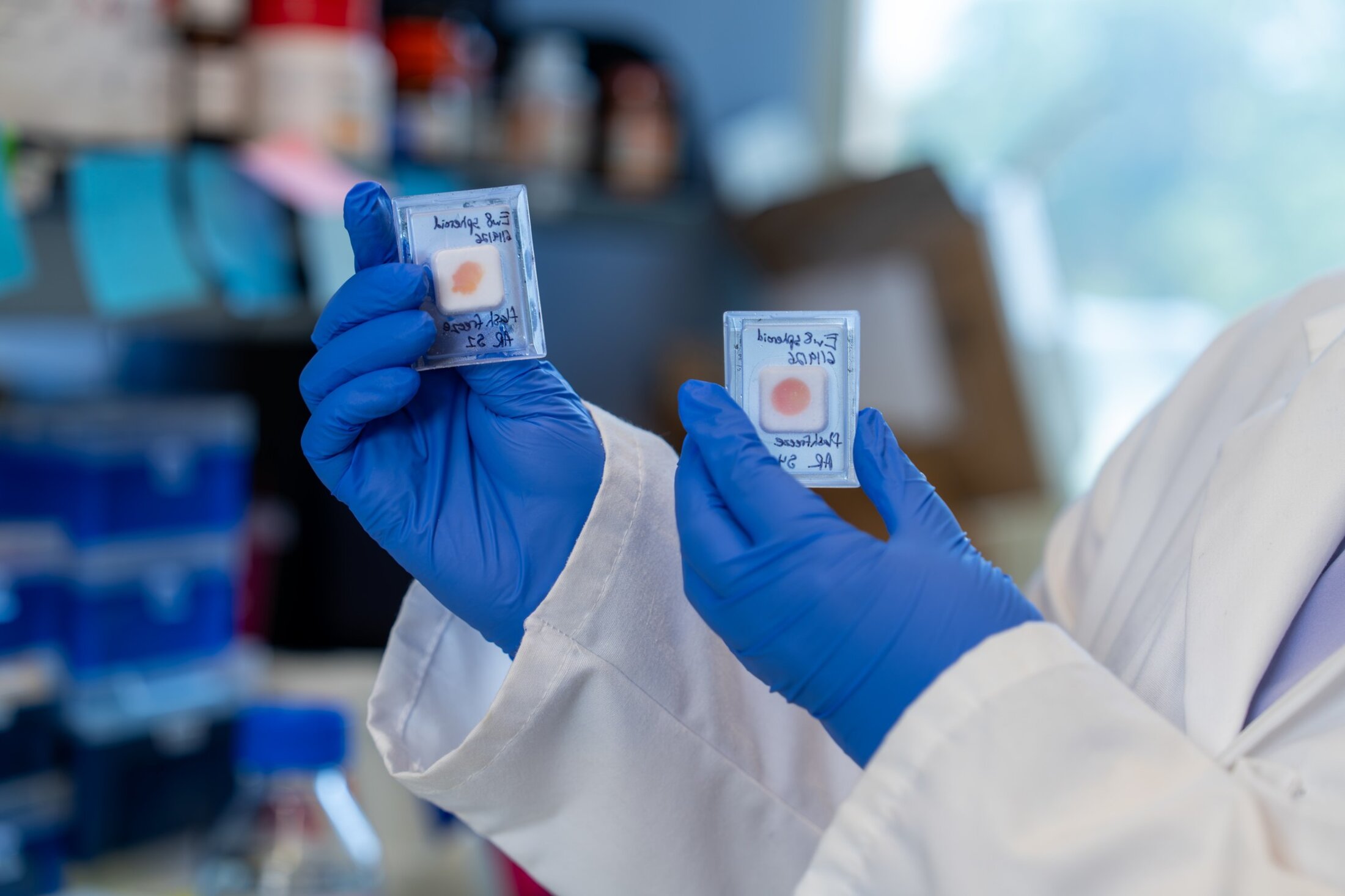
The resulting hydrogel, a creation whipped together with a precise ratio of ingredients and just the right amount of force applied to mixing, gives researchers the chance to understand how drugs act in an environment that more closely mimics real-body conditions.
So how does one make the intellectual leap from televised baking to cancer research?
Reno is a postdoctoral fellow in the lab of Casey Langdon, Ph.D., a pediatric sarcoma researcher at MUSC Hollings Cancer Center and the Darby Children’s Research Institute. As a bioengineer with a heavy materials science background, she focused her doctoral work at Clemson University on skin grafts.
Bioengineered skin grafts can be used as medical treatment for burns, for example, but they’re also used in research – to test cosmetics or to model skin problems like eczema or scleroderma. In fact, Reno continues to make models for MUSC rheumatologists and dermatologists to test drugs.
“They're just a great way, since animal skin is not very similar to human skin, for us to screen a drug and see how it will work on human patients,” Reno explained.
Similarly, although animal models are critical to many types of cancer research, they don’t work well for every cancer type. One of those types is bone sarcoma.
“There's no true genetic mouse model for Ewing sarcoma, which is one of the pediatric tumors with the least drug development in 40 years,” Reno said.
Ewing sarcoma of the bone is most often diagnosed in young people between the ages of 10 and 20. It's rare, affecting only about 200 young people each year. Similarly, osteosarcoma, another type of bone cancer, is most often diagnosed in teens. It affects about 1,000 people each year.
Traditional murine models haven’t worked well for studying these types of cancers, in part because human bones differ significantly in composition and material, making it difficult to reproduce how these cancers develop and spread in human bone accurately. Mice are notoriously able to squeeze through holes as small as a dime. That’s because the collagen in their bones is organized differently – it doesn’t have the mechanical integrity of human bone.
“It makes them very, very flexible. They can just squeeze into different spaces,” Reno said.
Humans, on the other hand, as larger creatures, need a lot more structural support.
“There's very different mechanical stiffness levels,” Reno explained. “And when you have different stiffness, cancer grows very differently. Cancer is more metastatic in a higher stiffness environment. So that's why we don't really see metastasis models in bone tumors in mice.”
Reno’s 3D models attempt to overcome this problem by more closely mimicking the makeup of the environment where sarcoma begins.
“A tumor is not just tumor cells – it has other cells. We’re not adding every single thing that is in your real tissue, but we are adding the base components. And because of that, we get a mechanical match where we can see drugs diffuse the same way they would in your body. We can see how your extracellular matrix, the stuff around your cells, interacts with the drug, not just the cells themselves,” Reno said.
That’s key because a drug that may work one on one against cancer cells in a Petri dish may not work as well in the body.
It’s especially important when testing drugs that could be used for children, who could live with the long-term side effects of cancer treatment for decades to come. For example, researchers would want to know if a drug could permanently affect bone density.
“That’s a really important thing because we don’t want to treat a child and then they have lifelong osteoporosis,” Reno said.
So far, the models are acting true to life. The cell signaling happening within them mirrors the signaling reported in the scientific literature, and they’re even starting to metastasize.
Models like this give us the ability to really fine-tune a drug and target it specifically to the tumor.
Reno said the lab is testing existing drugs that have already received Food and Drug Administration (FDA) approval for adults as well as new drugs developed in MUSC labs. The Dolloff Lab, for example, has provided a library of novel proteasome inhibitors to screen.
Reno has also, with the help of the Zucker Institute for Innovation Commercialization, patented the processes for creating these 3D models. She hopes to license the recipe to a company that would create kits to sell to research labs. The kits would make it easier for biology labs, most of which don’t have materials science expertise, to grow their own 3D models easily.
“They would just have to heat and warm some things instead of doing the whole process from powders,” Reno said. “There are already some kits like that on the market, but there are none for bone. There are very few for muscle and also very few for connective tissue.
“We ended up creating different ratios for skin, muscle, connective tissue and bone, and we patented all of those.”
Reno pointed out that these 3D models overcome one of the drawbacks of many other 3D models – size.
Most 3D models operate on a minuscule scale. They can recreate the complex workings of an organ, but studying these models is more akin to studying cells in a Petri dish. Reno’s models, though small to a nonscientist's eyes, are large enough to be frozen, sliced and viewed just like real tissue.
“That's where it kind of confuses some of the other 3D culture labs, because they're used to using small-scale techniques that are more similar to 2D culture, and they're like, ‘Whoa, you can actually freeze it and slice it?’ And I’m like, ‘Yes! You can!’”
There are benefits and limitations to both approaches, and both will be needed to continue to make progress against cancer. Reno would like to see more labs incorporating 3D models to get more accurate drug activity results before moving on to additional experiments.
“Models like this give us the ability to really fine-tune a drug and target it specifically to the tumor.”
Leslie Cantu
Senior Communications Manager
Leslie Cantu is the senior communications manager at MUSC Hollings Cancer Center, where she works with researchers, clinicians and patients to tell the people of South Carolina about the innovative work being done to improve cancer care for everyone in the state. She joined the MUSC Office of Communications and Marketing in 2018 after a career as an award-winning writer, editor and producer at community newspapers and local TV news. She transferred to the communications office at Hollings in 2022, where she happily finds something new and interesting to write about every day. Her favorite stories to cover at MUSC have included Match Day, the Angel Tree Parade, a clinical trial of CAR-T cell therapy and the many patients who have agreed to share their very personal struggles and triumphs.
Contact Leslie at cantul@musc.edu.