For surgeons removing cancer from the head and neck, every millimeter matters. Take too little tissue and cancer cells may be left behind. Take too much and surgery can affect structures essential for breathing, speaking, swallowing and other everyday functions.
A new clinical trial at MUSC Hollings Cancer Center is testing whether giving surgeons another way to see cancer – by making tumors glow – could help them to navigate that delicate balance.
The phase 2 pilot study, led by Jason Newman, M.D., director of the Division of Head and Neck Oncology, is evaluating a fluorescence imaging approach to highlight tumor tissue during surgery. The goal is to determine whether the added visual information helps surgeons to distinguish cancer more accurately from surrounding healthy tissue.
“The goal is safer surgery, a higher chance of negative margins and a better chance of leaving normal things behind because you’re not as worried about missing the tumor,” Newman said.
Finding the edge of a tumor
During cancer surgery, surgeons aim to remove the entire tumor along with a border, or margin, of healthy-looking tissue around it. A positive margin means cancer cells are present at the edge – an indication that some cancer may remain in the body. Positive margins are a risk factor for the cancer returning, but avoiding them can be particularly challenging in the head and neck.
We’re hopeful that a trial like this continues to push us closer to higher and higher levels of cancer care. If we can minimize the amount of tissue that we have to remove in order to achieve a better outcome, that combination is the sweet spot.
Unlike in some areas of the body where surgeons have more room to remove additional tissue around a tumor, head and neck tumors can sit close to the tongue, throat, voice box and major blood vessels and nerves involved in movement, breathing and swallowing. Removing additional tissue can come with significant consequences.
“Your decisions during surgery have to be precise, or else you start creating new negative side effects,” Newman said.
Surgeons traditionally rely largely on what they can see under normal white light, along with what they can feel and what they know about the tumor from scans and other information collected before surgery. But the boundary between cancer and healthy tissue is not always obvious to the naked eye.
That is where molecular imaging adds another layer of information.
Making cancer light up
Surgeons in the new trial are taking advantage of the different way a fluorescent dye called indocyanine green, or ICG, behaves in cancerous versus healthy tissue. After the dye is injected, it gradually clears from most healthy tissue but remains in the tumor. That creates a period known as the “second window” when the tumor contains more dye than the surrounding tissue.
“You’re left with a tumor that glows, but everything else around it is not glowing anymore,” Newman said. “That gives the surgeon another piece of information to help determine where the tumor ends and where it is safe to cut.”


Patients in the trial receive the ICG dye through an IV up to five days before surgery. During the operation, surgeons use a special light and camera to detect the dye, making the tumor appear to glow on a screen. They can view the tumor in real time and then check the surgical area afterward to see whether any glowing tissue – a sign of remaining cancer – is left behind.
The technology is designed to complement, rather than replace, the surgeon’s usual view. Surgeons can toggle between what they see under normal light and the fluorescence image, using the additional information as needed throughout the operation. The goal is for the technology to fit seamlessly into the normal surgical workflow, giving surgeons another tool to guide their decisions without fundamentally changing how the operation is performed.
Putting the glow to the test
Seeing a tumor glow is compelling. But the purpose of the trial is to determine whether that glow provides surgeons with practical information. One of the central questions is whether fluorescence changes where a surgeon believes the tumor ends and whether it leads to more accurate decisions about how much tissue to remove.
“These are the hypotheses of this clinical trial,” Newman emphasized. “We haven’t proved that this improves surgery yet – that’s why we’re doing the trial. We need to objectively demonstrate that this additional information makes a difference.”
Researchers are enrolling 30 adults with head and neck squamous cell carcinoma who are undergoing surgery and at a higher risk of cancer recurrence.
Researchers will compare what surgeons see under traditional white light with what they see using ICG fluorescence. They will then compare both assessments with the resulting pathology – the microscopic examination of the removed tissue that remains the gold standard for determining whether cancer cells are present.
If the pilot study produces encouraging results, Newman said the next step would be to test the approach in a larger group of patients and across multiple surgeons and institutions.
Building the future of cancer surgery
For Newman, the trial represents more than a single experiment in head and neck cancer.
He and Dauren Adilbay, M.D., Ph.D., a Hollings head and neck surgeon with expertise in molecular imaging, are working to build a broader program around fluorescence-guided surgery at MUSC. For instance, head and neck surgeons have tested a fluorescent dye to illuminate nerves during surgery, with the goal of better identifying and preserving critical nerves. Adilbay is also developing a fluorescence-based approach to predict precancerous oral lesions most likely to develop into cancer.
Ultimately, Newman envisions researchers identifying imaging agents and molecular targets that could allow surgeons to visualize a variety of cancers more effectively.
“This is more like starting to lay a foundation,” Newman said.
That foundation could eventually extend beyond head and neck cancer to surgical specialties treating breast, colorectal, gynecologic and other cancers, he said.
The current investigator-initiated study was supported through internal Hollings funding, which Newman said provided the early investment needed to test a promising idea before enough evidence exists to compete for larger external grants. The hope is that data generated through the pilot trial can support larger studies evaluating whether fluorescence-guided surgery improves outcomes.
For patients with head and neck cancer, the long-term vision is straightforward: Give surgeons more information to remove the cancer while preserving as much healthy tissue – and function – as possible.
“We’re hopeful that a trial like this continues to push us closer to higher and higher levels of cancer care,” Newman said. “If we can minimize the amount of tissue that we have to remove in order to achieve a better outcome, that combination is the sweet spot.”
Featured in this story
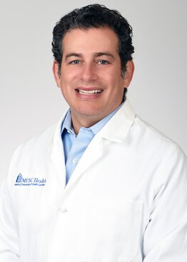
Jason Newman, M.D., FACS
As a physician providing cancer care daily to people who come to Charleston from across the state, Jason Newman, M.D., believes deeply in the importance of the strategic growth of cancer programs at Hollings Cancer Center. The expansion of clinical care, research and clinical trials makes innovative treatments more accessible to more patients and helps Hollings reach populations in rural and underserved areas. As the network grows, regional teams will be able to deliver care to patients locally with specialized treatment available in Charleston. In his role as the Oncology Integrated Center of Clinical Excellence (ICCE) chief, Newman oversees the clinical activities and performance of Hollings and executes strategies for program development and growth.

Hayley Kamin
Communications Manager
Hayley Kamin is the communications manager for the Hollings Cancer Center Communications and Marketing team, having joined the team in 2025 after three years as a communications specialist at the National Institutes of Health (NIH). As a science communicator with a Ph.D. from the University of Florida, she has extensive experience translating complex research into clear, engaging content. Her career has included roles at the NIH’s National Institute of Mental Health and the American Psychological Association, where she led content development and editorial strategy, developed science and health communications and worked with researchers and clinicians to strengthen public understanding of research.
Contact Hayley at kamin@musc.edu.
Recent Cancer Clinical Trials stories