Services
- Access to state-of-the-art confocal and multiphoton microscopy, super-resolution microscopy, and second harmonic generation for imaging living cells in culture, live, and fixed tissues and for intravital imaging in live animals.
- In-depth training in multiple imaging modalities including microscopic techniques, live cell imaging, multiphoton imaging, and pre-clinical intravital animal imaging.
- Consultation and assistance concerning experimental design, sample preparation, probe selection, and data analysis for imaging applications.
- Education in the fundamentals of imaging and image analysis technologies and their application, which are essential for appropriate utilization of these approaches.
- Dissemination of information concerning the ongoing development of novel experimental strategies, emerging technologies, and data analysis techniques.
Applications
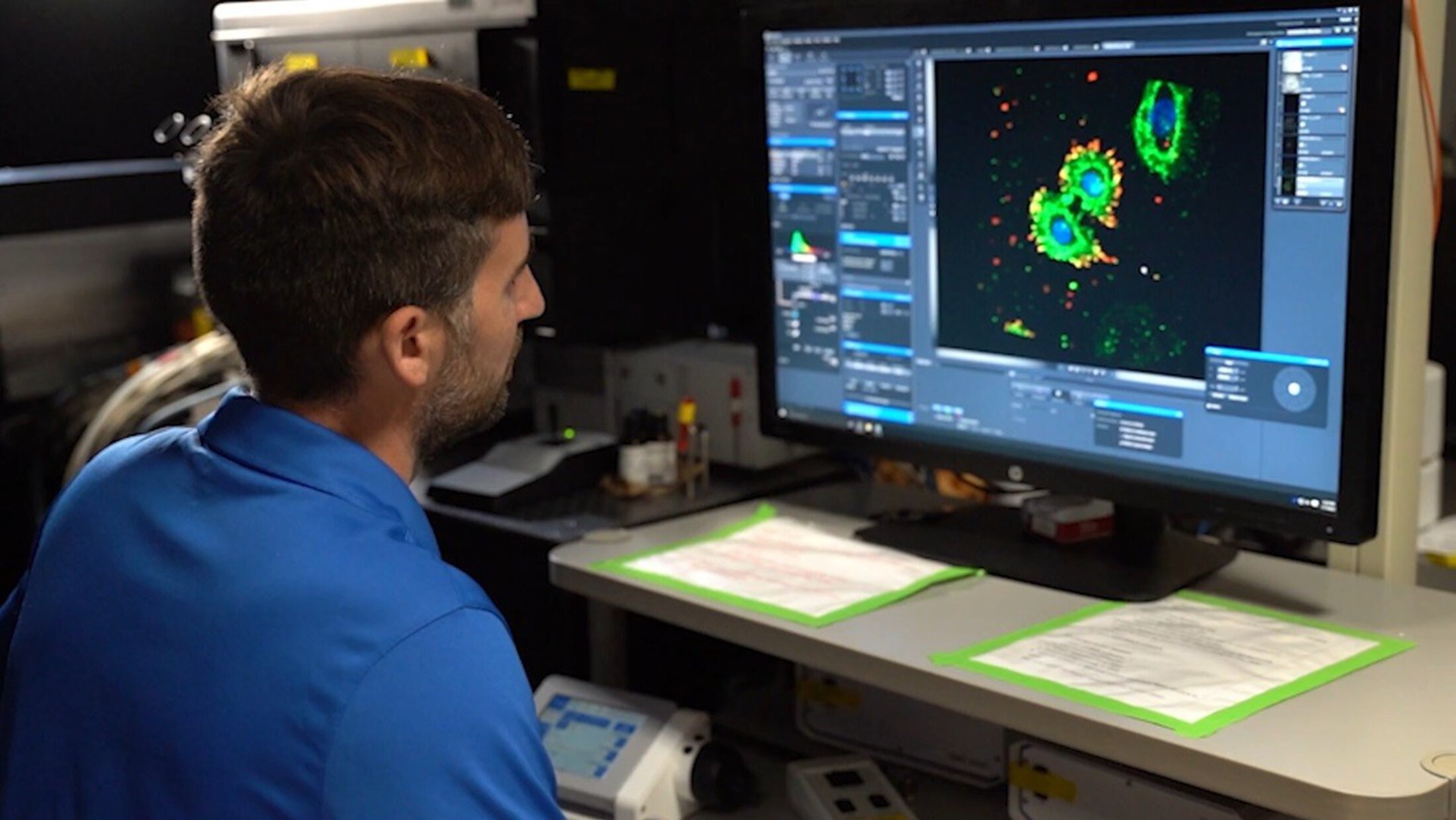
Fixed Cell & Tissue Imaging using chromogenic and fluorescence techniques in combination with antibody-based methods to monitor expression, distribution, and interaction of specific molecules.
Live Cell Imaging of parameter-sensitive fluorophores to monitor ions, electrical potentials, oxygen and nitrogen radical generation, NAD(P)H, mitochondrial and plasmalemmal membrane permeability, cell viability (apoptosis and necrosis), fluorescent protein biosensors, and other parameters.
Intravital Microscopy to monitor microcirculation, leukocyte margination, invadopodia, mitochondrial polarization, membrane permeability, radical generation, gene expression, detection of collagen fibers, and other factors in living animals and tissues.
Advanced Imaging Techniques Fluorescence resonance energy transfer (FRET) and fluorescence recovery after photobleaching (FRAP) to characterize and quantify interactions between specific molecules and their mobility; second harmonic generation (SHG) microscope for label-free visualization of collagen; and Fast Airyscan super-resolution imaging with high quantum efficiency GaAsP photomultipliers.
Equipment
The CMI Shared Resource has a full range of state-of-the-art imaging equipment, including the microscopes listed below. Check out our equipment page for full microscope descriptions and capabilities.
- Leica Stellaris 8 TauSTED Xtend Confocal/Super-resolution.
- Zeiss LSM 880 NLO Confocal/Multiphoton with Fast Airyscan Super-resolution Detector.
- Olympus FV1200 MPE.
- Andor BC43 Spinning Disk Confocal.
- Zeiss LSM 510 META.
- Olympus FluoView FV10i LIV.
- BD Biosciences CARV II.
Imaging Workstation
Two imaging workstations are dedicated for offline image processing and analysis. The following software packages are available for current CMI users: Metamorph, Image J FIJI, Duolink ImageTool, Adobe Photoshop, Olympus Viewer, IP Lab, Zeiss Zen software, Imaris, and Huygens deconvolution suite. Please contact Li Li, Ph.D., for more information and to request software training.
Additional Equipment for Live Cell Imaging
A tissue culture hood and two cell culture incubators (37°C) are available in Drug Discovery 521 for specimen preparation and temporary incubation of cells. One incubator is equipped for 5% CO2.
Publication Acknowledgment
Please acknowledge the Cell & Molecular Imaging Shared Resource in your publications by choosing the appropriate grant numbers: “Image facilities were supported in part by the Cell & Molecular Imaging Shared Resource, MUSC Cancer Center Support Grant (P30 CA138313), the SC COBRE in Oxidants, Redox Balance, and Stress Signaling (P20 GM103542), the SC COBRE in Digestive and Liver Diseases (P20 GM130457), the MUSC Digestive Disease Research Cores Center (P30 DK123704,) and the Shared Instrumentation Grants S10 OD018113 and S10 OD028663.”